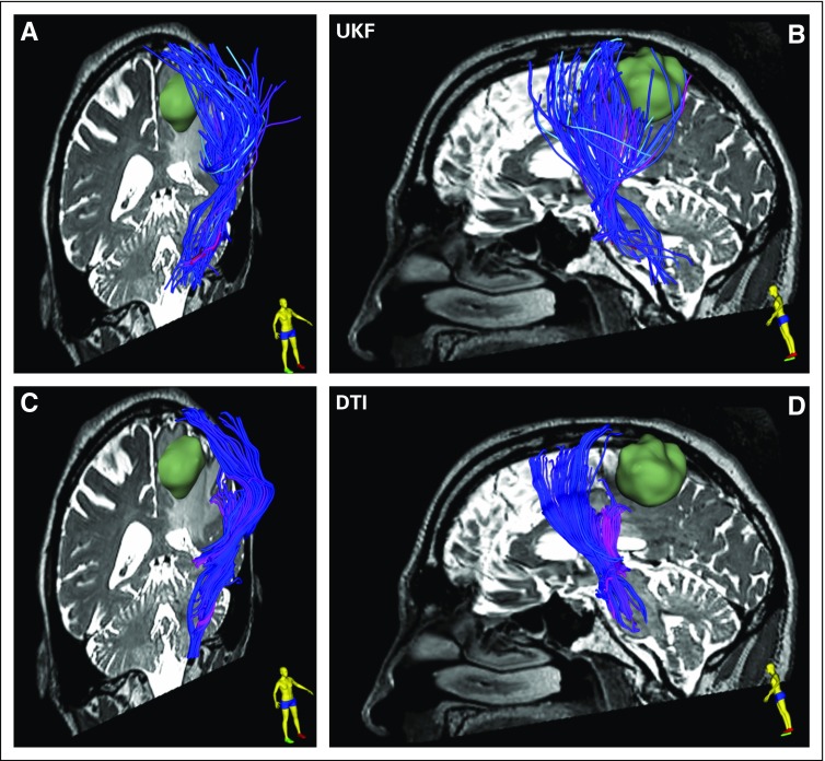
FIG 3.
Single-tensor diffusion tensor imaging (DTI) v two-tensor unscented Kalman filter (UKF) tractography with a free water model of edema in patient data set 1. The tracts are visualized using tubes in three dimensions, with a diameter of 1 mm and colored by mean fiber orientation. (A and C) An oblique view from the right, featuring a coronal image, tumor in green, and corticospinal tract (CST) anterior to the tumor. The lateral projections of the CST that cross through edema and intersect with long anterior-posterior fibers are difficult to trace with conventional single-tensor DTI but can be traced using two-tensor UKF tractography. (B and D) A sagittal view of the relationship of CST and tumor on the basis of the two tractography methods. The UKF method with the free water model traces a seemingly larger volume of fibers within the edema that surrounds the tumor.

