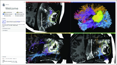
FIG 6.
The 3D Slicer user interface that shows visualization of navigated intraoperative ultrasound (iUS) overlaid on preoperative T1-weighted magnetic resonance imaging with contrast along with the automatically identified language fiber tracts in patient data set 2 (tracts and colors as in Fig 4). The iUS image is used retrospectively here for a demonstration of software functionality. iUS information demonstrates brain shift that occurs after dural opening and likely displaces the arcuate fasciculus (purple), which can be seen at the edge of various parts of the resection cavity in each panel (top left, axial; bottom left, sagittal; bottom right, coronal). By integrating this brain shift information with tractography, the surgeon can make an updated estimate of how far the tracts might be. DICOM, Digital Imaging and Communications in Medicine.

