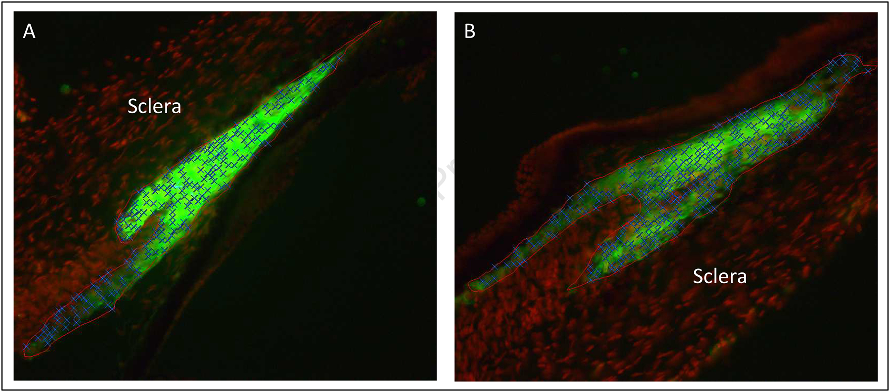
Figure 2: Representative Post-Treatment Stained Ciliary Muscle Images (20x objective).

Images are from the treated (A) and fellow (B) eyes of the same animal. The ciliary muscle has been outlined in red, smooth muscle fibers have been stained green (anti-smooth muscle actin conjugated with FITC), and cell nuclei have been stained red (Draq5). Note that ciliary muscle cell nuclei have been labeled with blue Xs.
