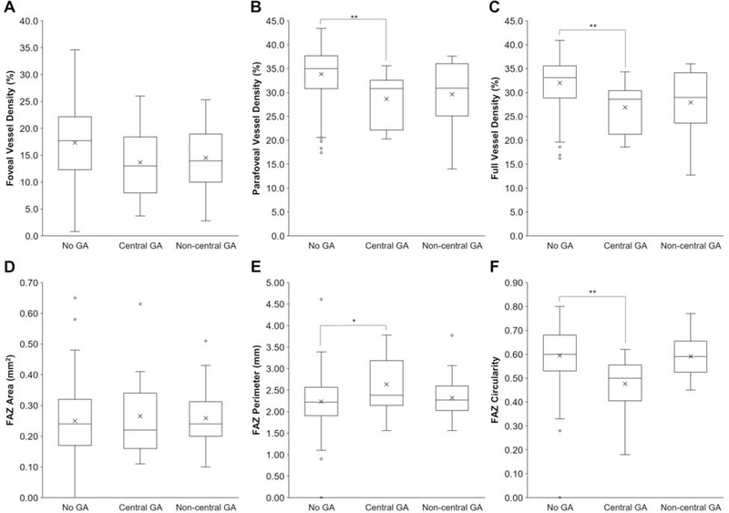
Figure 5. Retinal Vascular Measurements in Geographic Atrophy.
Box-and-whisker plots comparing foveal (A), parafoveal (B), and full (C) VD, as well as FAZ area (D), perimeter (E), and circularity (F) in eyes with non-exudative AMD and no geographic atrophy (GA), central GA, or non-central GA. *, P<0.05; **, P<0.01

