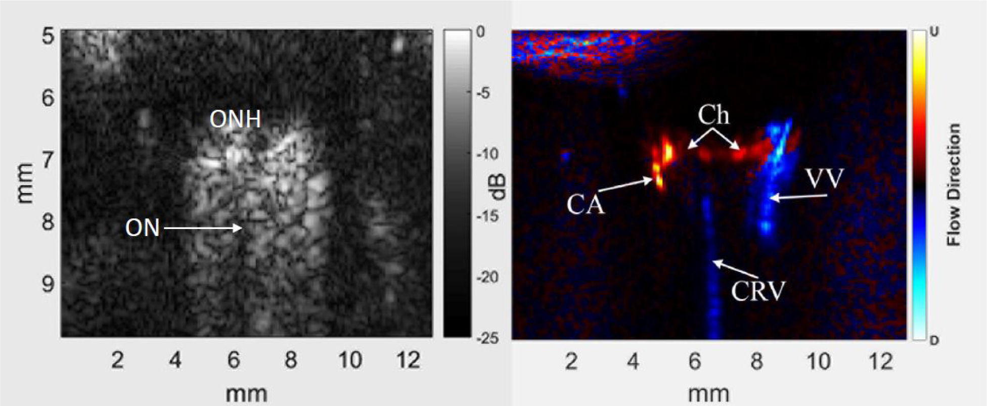
Figure 2.

Representative B-scan (left) and power Doppler image (right) of the posterior pole and retrobulbar region of a rat eye before injection of contrast. The optic nerve (ON) appears as a non-reflective region extending posteriorly from the optic nerve head (ONH) into the orbital tissue. Colors in the power Doppler image depict flow direction: red towards the probe and blue away, usually indicative of arterial and venous flow, respectively. In the power Doppler image, the central retinal vein (CRV) and a vortex vein (VV) are evident, as is choroidal flow (Ch) and a long posterior ciliary artery (CA). Note that images are ‘stretched’ axially.
