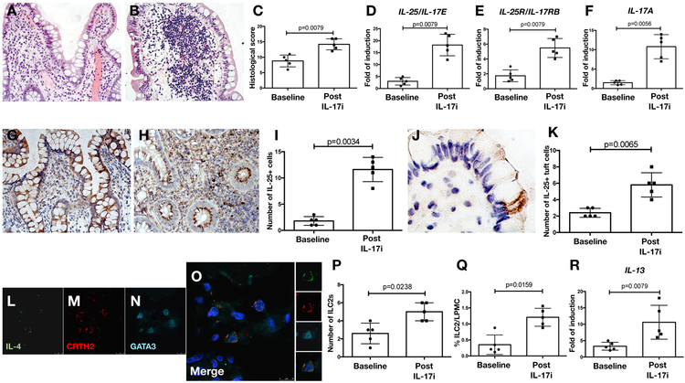
Figure 6: IL-25 driven inflammation characterizes colitis seen in in SpA (AS) subjects after IL-17i therapy.
A-B: Representative hematoxylin and eosin staining images demonstrating histologic changes in the ileum of SpA (AS) subjects at baseline (A) and after the onset of clinically evident Crohn’s disease (CD) (B) during treatment with IL-17i (secukinumab). C: Histological score in SpA (AS) ileal samples at baseline and after the onset of CD. D-F: Relative mRNA levels of IL-25/IL-17E (D), IL-25R/IL-17RB (E) and IL-17A (F) in five SpA (AS) subjects at baseline and after the onset of CD as assessed by RT-PCR. G-H: Representative images showing IL-25 immunostaining in SpA (AS) ileal samples at baseline and after the onset of CD. I: Number of IL-25 positive cells the gut of SpA (AS) subjects. J: Representative image showing the expression of IL-25/IL-17E in the context of specialized epithelial cells that were morphologically identified as tuft cells in the gut of SpA (AS) subjects after the onset of CD during treatment with IL-17i (secukinumab). K: Number of IL-25 positive tuft cells at baseline and after the onset of CD. L-O: Representative confocal microscopy images of IL-4 (L), CRTH2 (M) and GATA-3 (N) in SpA (AS) ileal tissue after the onset of CD during treatment with IL-17i (secukinumab). O: Merge triple staining showing IL-4/CRTH2/GATA3 positive ILC2 co-localization in the gut of AS (SpA) patients. P: Number of type-2 innate lymphoid cells (ILC2s) in the gut of SpA (AS) subjects. Q: Percentage of ILC2s evaluated by flow cytometry in the gut of SpA (AS) subjects at baseline and after the onset of CD during IL-17i (secukinumab) treatment. R: Relative mRNA levels of IL-13 in the ileum of five SpA (AS) subjects at baseline and after the onset of CD as assessed by RT-PCR. Statistical comparisons calculated using the Student’s t-test and the Mann-Whitney U test.

