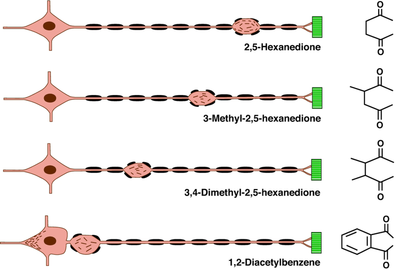
Figure 2.
Nerve fiber response to γ-diketones. Diagram of the location of initial focal axon (orange) swellings and associated secondary myelin (black) retraction in an elongate, large-diameter peripheral nerve fiber originating from the neuron soma (left) and terminating at sites of muscle (green) innervation. γ-Diketone potency and associated neuroprotein reactivity increase from 2,5-hexanedione (2,5-HD, upper) to 1,2-diacetylbenzene (1,2-DAB, lower), where neurofilaments accumulate both in the neuronal soma, distal to the initial segment, and in proximal spinal roots (as shown). The differential spatial “rigidity” (ability to rotate in space) of the 1,4-diketo groups may dictate the protein reactivity and, hence, neurotoxic potency of the illustrated γ-diketones. This diagram illustrates the principle that the toxic potential of the γ -diketone dictates how far neurofilaments are transported from the neuron cell body along the nerve fiber before they accumulate distally in focal axonal swellings; the greatest transport distance representing the least potent (2,5-HD) and, hence, the slowest developing effect. The most potent γ -diketone, 1,2-DAB, rapidly reacts with axonal proteins, such that anterograde transport of neurofilaments is arrested before or shortly after their departure from the neuron cell body. This figure is constructed from the author’s examination of single teased nerve fibers, and light and electron micrographs of cross-sectioned nerves, from peripheral nerve fibers undergoing γ -diketone axonopathy [2,51,52,59,60,62,108,113,114].

