FIGURE 1.

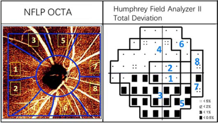
In a perimetric glaucomatous eye, the 4.5x4.5-mm en face OCT angiogram of the nerve fiber layer plexus (NFLP) and the 24–2 visual field (VF) map were divided into 8 corresponding sectors according to a modified Garway-Heath scheme.

In a perimetric glaucomatous eye, the 4.5x4.5-mm en face OCT angiogram of the nerve fiber layer plexus (NFLP) and the 24–2 visual field (VF) map were divided into 8 corresponding sectors according to a modified Garway-Heath scheme.