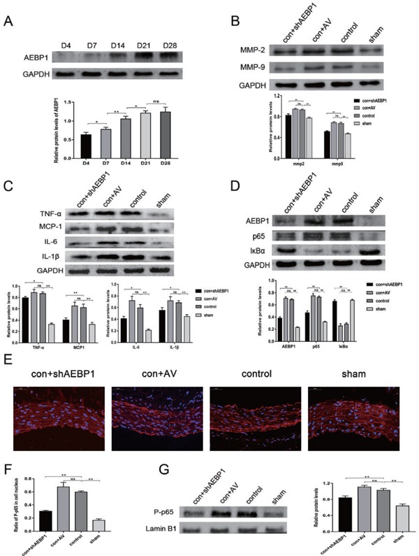
Fig. 5.

Expression profiles of AEBP1, the NF-κB pathway factors, inflammatory cytokines, and MMPs in the established AAA model (n = 10)
(A) AEBP1 expression was measured using the western blot at days 4, 7, 14, 21, and 28 (n = 3) after the surgery. (B) MMP2 and MMP9 expression were evaluated using the western blot (with GAPDH as the loading control). (C) Evaluation of the levels of inflammatory cytokines TNFα, MCP-1, IL-6, and IL-1β by the western blot. (D) Protein levels of AEBP1, NF-κB p65, and IκBα were evaluated by western blot in the established AAA model. (E) Immunofluorescence staining and micrographs (400X) indicated P-p65 expression in the infrarenal abdominal aorta in different groups (n = 4). (F) Quantification of the proportion of P-p65 positive nuclei. (G) Nuclear proteins were extracted from the infrarenal abdominal aorta (n = 4), and P-p65 expression was evaluated using the western blot. All the relative expression levels are presented as mean ± SD; one-way ANOVA, ns > 0.05,*p < 0.05; **p < 0.01. GAPDH was used as the loading control in C and D, whereas Lamin B1 was used in G.
