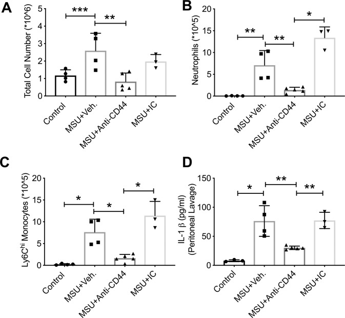
Figure 6.
Impact of anti-CD44 antibody treatment on inflammatory cell infiltration and production of interleukin-1 beta (IL-1β) in murine peritoneal monosodium urate monohydrate (MSU) crystal inflammation model. MSU crystals (2 mg in 200 μL PBS), vehicle (Veh.; 200 μL PBS), anti-CD44 and isotype control (IC) antibodies (50 μg in 200 μL PBS) were administered via the intraperitoneal route. Experimental groups included untreated controls (n = 4), MSU + Veh. (n = 4), MSU + Anti-CD44 antibody (n = 5) and MSU + IC antibody (n = 3). Peritoneal lavaging was performed at 4 hours. Total peritoneal lavage cell counts were determined. The number of infiltrated neutrophils and monocytes were determined using flow cytometry and probing for neutrophil markers; Ly6B.2 and Ly6G and monocyte markers; Cd11b and Ly6C. Representative flow cytometry plots of neutrophil and monocytes markers are shown in Supplementary Fig. 4. Peritoneal lavage IL-1β levels were determined by ELISA. *p < 0.001; **p < 0.01; ***p < 0.05. (a) Anti-CD44 antibody treatment reduced the number of cells in peritoneal lavages following MSU administration. (b) Anti-CD44 antibody treatment reduced neutrophil infiltration in MSU peritoneal inflammation model. (c) Anti-CD44 antibody treatment reduced Ly6Chi monocyte infiltration in MSU peritoneal inflammation model. (d) Anti-CD44 antibody treatment reduced lavage IL-1β levels compared to vehicle or IC antibody treatments.

