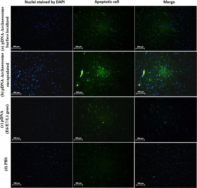
Figure 5.
Tumor cell apoptosis in vaccinated mice. For the analysis of the apoptosis marker in vivo, TC-1 tumor-bearing animals were vaccinated with each vaccine groups or PBS. Tumor apoptosis was detected using TUNEL and analysed by fluorescence microscopy in (a) pDNA-surface localized archaeosomes group, (b) pDNA-encapsulated archaeosomes group, (c) L1/E6/E7 recombinant gene alone group and (d) PBS control group. Total nuclei are visualized in blue (DAPI) and TUNEL-positive nuclei are stained in green (FITC). The images are representative of three independent experiments. TUNEL analysis indicated that the combined L1/E6/E7 + archaeosomes treatment was associated with more apoptosis (40–60%) than the L1/E6/E7 alone (15–20%). Controls (no treatment) showed a low background level of apoptosis (0–1.5%). All images are shown at × 40 magnification.

