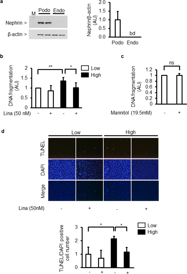
Figure 1.
Effect of high glucose and linagliptin on apoptosis in cultured podocytes. (a) Immunoblot analyses of nephrin in cultured podocytes and endothelial cells. Podo; podocytes. Endo; endothelial cells. bd; below detection limit. (b) DNA fragmentation in podocytes incubated with low glucose (5.5 mM) or high glucose (25 mM) for 96 h in the absence or presence of linagliptin (50 nM). Lina; linagliptin. *P < 0.05. **P < 0.01. (c) Effect of D-mannitol-induced hyperosmolality on DNA fragmentation. Low; low glucose. High; high glucose. Lina; linagliptin. ns; not significant. (d) Immunocytochemical staining with TdT-mediated dUTP nick end (TUNEL) and 4′,6-diamino-2-phenylindole (DAPI) (merged image). Magnification: X40. Low; low glucose. High; high glucose. Lina; linagliptin. *P < 0.05. These data are expressed as means ± SD. Results are representative of one of three independent experiments.

