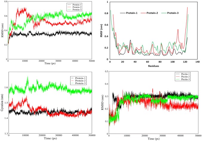
Fig. 5.
RMSD, RMSF and Gyration analysis. (Upper left) RMSD values of test proteins showing the structural stability of Celosia cristata DUF538 protein as protein 1 in comparision to C-terminus of Brassica napus pectin metylesterase as protein 2 and Arabidopsis thaliana pectinesterase as protein 3 during simulation time. (Upper right) RMSF values of test proteins showing the structural stability of Celosia cristata DUF538 protein as protein 1. (Lower left) Gyration values of test proteins showing the structural stability of Celosia cristata DUF538 protein as protein 1. (Lower right) RMSD values of ligand showing the stability of DUF538-pectin interactive structure as compared to others

