FIGURE 3.
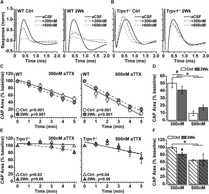
(A,B) Example compound action potential (CAP) responses of nerves from Ctrl eyes and following 2 weeks of elevated intraocular pressure (IOP) from wild-type (WT) and Trpv1–/– mice with bath application of 300 and 600 nM of aTTX. (C) Mean WT CAP area for control and 2-week nerves decreases over time following bath application of 300 and 600 nM of aTTX. Individual recordings normalized to corresponding baseline (pre-drug) response. Slopes of best-fitting regression lines indicated significant decline (p-values indicated). (D) Final CAP area for WT decreases significantly following 300 nM of aTTX for both control (n = 7, 50% decrease) and 2-week (n = 6, 59% decrease) nerves compared with baseline for each (#p ≤ 0.03). CAP area decreased further from baseline for control (n = 6, 91% decrease) and 2-week nerves (n = 6, 83% decrease) following application of 600 nM of aTTX, both significant declines compared with 300 nM (*p < 0.001). (E) Mean Trpv1–/– CAP area following bath application of 300 and 600 nM of aTTX; for slopes of best-fitting regression lines, only control nerves with 600 nM of aTTX showed significant decline (p-values indicated). (F) Final CAP area for Trpv1–/– control nerves were minimally affected by 300 nM of aTTX (n = 5, 0.5% decrease), whereas area for 2-week nerves declined compared with baseline (n = 5, 18% decrease; #p = 0.02). Like WT, 600 nM of aTTX caused a greater reduction in CAP area compared with 300 nM for Ctrl nerves (35% decrease; *p = 0.02). Statistics: (C,E) linear regressions; (D,F): one-way ANOVAs, Tukey post-hoc.

