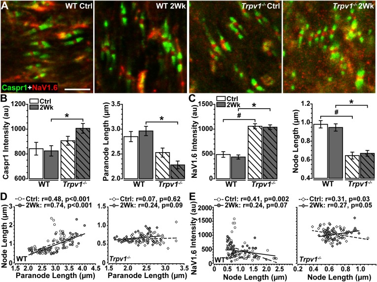
FIGURE 5.
(A) High magnification confocal micrographs of longitudinal sections through Ctrl and 2 Wk wild-type (WT) and Trpv1–/– optic nerves show Caspr1-labeled paranodes (green) flanking NaV1.6 (red) within nodes of Ranvier. (B) Trpv1–/– 2 Wk paranodes contain increased Caspr1 than did WT 2 Wk nerves (left, *p = 0.012) and are shorter (right, *p < 0.001). (C) NaV1.6 is higher in nodes of Ranvier of Trpv1–/– nerves (left) compared with WT Ctrl (#p < 0.001) and 2 Wk (*p < 0.001) optic nerve (left), although node length is significantly shorter for both Trpv1–/– Ctrl (#p < 0.001) and 2 Wk (*p < 0.001) nerves. Elevated intraocular pressure (IOP) had no effect on either measure (p ≥ 0.88). (D) There is a positive relationship between node and paranode lengths in WT optic nerves (left), however, this relationship is lost in Trpv1–/– (right) optic nerves. Elevated IOP had no effect on this relationship for WT or Trpv1–/– optic nerves. (E) NaV1.6 intensity decreases as node length increases in WT (left) and Trpv1–/– (right) control optic nerves. Following IOP elevation, this relationship is lost in WT nerves, whereas the relationship becomes positive in Trpv1–/– nerves. Scale = 5 μm (A). Statistics: (B,C): one-way ANOVAs, Tukey post-hoc; (D,E) linear regressions. Total nodes analyzed: WT Ctrl, 3,942; WT 2 Wk, 3,890; Trpv1–/– Ctrl, 2,024; Trpv1–/– 2 Wk, 2,191. Five animals per condition. Ten images per animal.

