Figure 1.

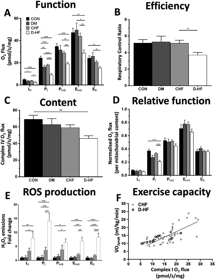
Mitochondrial function is impaired in the skeletal muscle of patients with D‐HF. Oxygen flux in all respiratory states (A) and the mitochondrial coupling efficiency as indicated by the respiratory control ratio (RCR) (B) is lower in D‐HF patients compared with DM and CHF. Mitochondrial content (measured by complex IV activity) is the lowest in D‐HF patients (C), and impairments at complex I remain despite normalizing for the lower mitochondrial content (D). These impairments corresponded to higher concentrations of mitochondrialderived reactive oxygen species (ROS) across all respiratory states in patients with D‐HF (E). N = 25, 10, 52, and 28 for CON, DM, CHF, and D‐HF, respectively. *P < 0.05; **P < 0.01; ***P < 0.001; ****P < 0.0001. Complex I function was strongly correlated to VO2peak as a measure of whole‐body exercise capacity in both patients with CHF (R2 = 0.47; P < 0.001; solid line; N = 34) and even more so in D‐HF (R2 = 0.64; P < 0.001; dashed line; N = 15) (F). EI + II, maximal uncoupled complex I + II respiration; EII, uncoupled complex II respiration; LI, complex I leak respiration; PI, complex I oxidative phosphorylation; PI + II, complex I + II oxidative phosphorylation.
