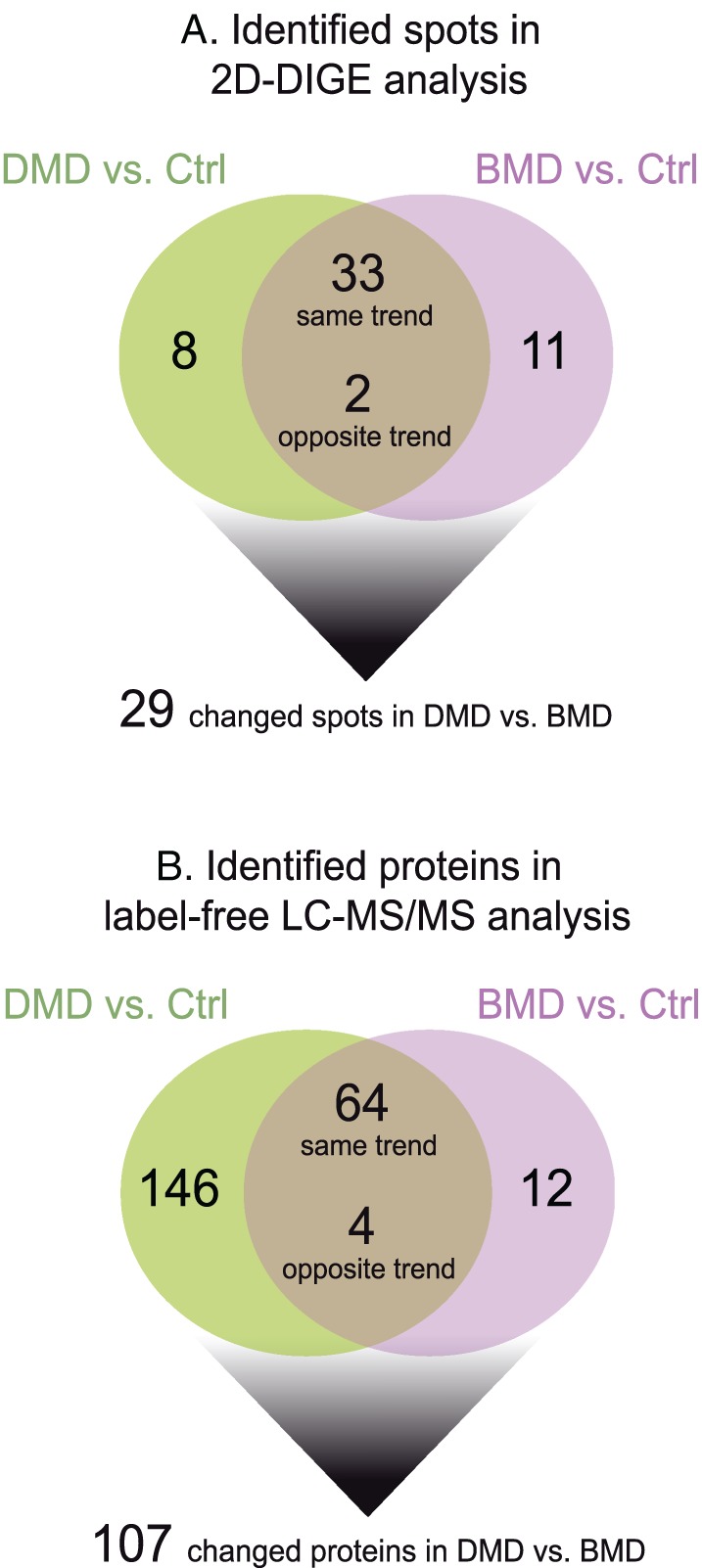
Figure 1.

Schematic diagrams resuming findings obtained from (A) 2D‐DIGE and (B) label‐free LC‐MS/MS proteomic analyses. 2D‐DIGE, two‐dimensional difference in gel electrophoresis; BMD, Becker muscular dystrophy; DMD, Duchenne muscular dystrophy; LC‐MS/MS, liquid chromatography with tandem mass spectrometry.
