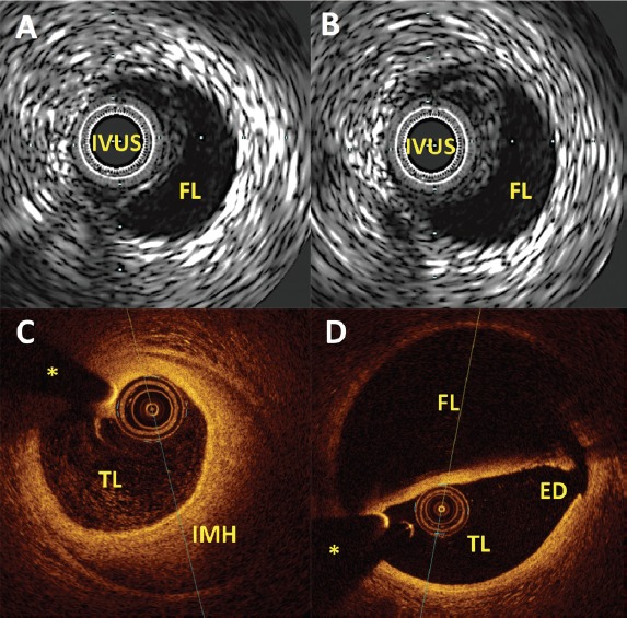
Figure 3: Intracoronary Imaging.

A and B: IVUS images in spontaneous coronary artery dissection, confirming the presence of the IVUS catheter within the true lumen and clearly depicting the presence of an anechoic FL; C and D: Optical coherence tomography images in spontaneous coronary artery dissection; C clearly depicts the presence of the catheter within the TL with and near 180° IMH. D: Optical coherence tomography high spatial resolution enables the clear definition of the ED of the dissection. ED = entry door; FL = false lumen; IMH = intramural haematoma; IVUS = intravascular ultrasound; TL = true lumen. *Wire artefact.
