
Fig. 1.
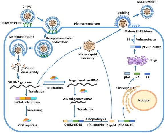
Schematic representation of the replication cycle of chikungunya virus. CHIKV enters the cell by endocytosis following the binding of the E2 protein to specific receptor(s) on the cell surface. Within the endosome, the low pH triggers the fusion of the viral envelope with the endosomal membrane, leading to the release of the nucleocapsid into the cytoplasm. The nucleocapsid disassembles to liberate the viral genome, which is translated to produce the viral nonstructural proteins (nsP1–4). After processing, the nonstructural proteins complex to form the viral replicase, which catalyzes the synthesis of a negative-sense RNA strand to serve as a template for synthesis of both the full-length positive-sense genome and the subgenomic (26S) RNA. The subgenomic (26S) RNA is translated to produce the structural polyprotein (C-E3-E2-6K-E1), which is then cleaved to produce the individual structural proteins, followed by assembly of the viral components. The assembled virus particle is released by budding through the plasma membrane, where it acquires the envelope with embedded viral glycoproteins.