
Fig. 5.
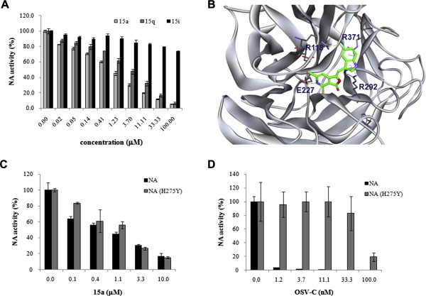
Inhibition of influenza virus NA activity by 15a and 15q. (A) NA activity assay. The enzymatic activity of the PR8 NA protein was measured in the presence of increasing amounts of 15a, 15q, or 15i using a chemiluminescent NA substrate. The data shown are the means ± S.D. for two samples from three independent experiments. (B) Docking analysis of 15a with the PR8 NA protein. The dashed lines represent interactions between the ligand and the target protein. The NA inhibitory molecule 15a is shown as a stick model, and the atom color-coding is as follows: carbon, green; nitrogen, blue; oxygen, red; sulfur, yellow; and fluorine, cyan. Inhibition assay of the purified wild-type and H275Y mutant NA proteins by 15a (C) and OSV-C (D). These proteins are derived from influenza H1N1 (A/California/04/2009) and its OSV-resistant H275Y mutant [NA (H275Y)]. Each experiment was performed independently twice in duplicate. (For interpretation of the references to color in this figure legend, the reader is referred to the web version of this article.)