
Fig. 4.
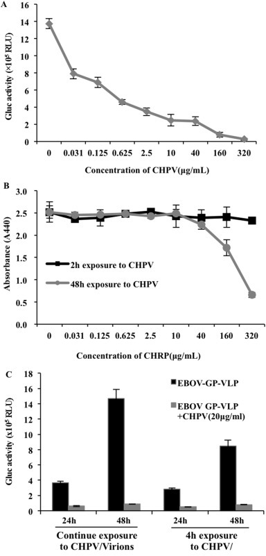
Evaluating the anti-EBOV effect and cytotoxicity of CHPV in vascular endothelial cells (HUVECs) and its anti-EBOV effect in macrophages. (A) The same amount of pseudovirions mixed with the indicated varying concentrations of CHPV were immediately added onto HUVECs for 2 h, then the cells were washed and cultured in DMEM without CHPV. The Gluc activities were tested from the supernatants after 48 h of incubation. (B) The cells were treated with CHPV for 2 or 48 h, after which the WST-1 cell proliferation assay was used to analyze cell proliferation. (C) Left 4 bars: CHPV was added into the macrophage culture 12 h before infection when EBOV-GP-V was added and continually cultured without washing; right 4 bars: EBOV-GP-V alone or mixed with CHPV (20 ug/ml) and immediately added onto cell culture, followed by washing of the cells after 4 h of incubation. The cells were then cultured in medium without CHPV. The supernatants of all samples were collected at 24 or 48 h post-infection and used to detect Gluc activity.