
Figure 1.
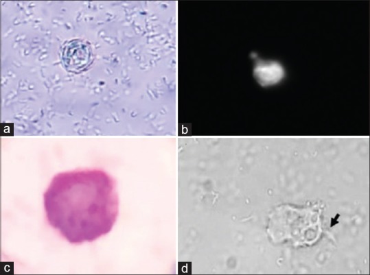
Acanthamoeba cyst and trophozoite stages in CSF cultures from reported cases: (a) wet mount (400×) (b) calcofluor stain (400×) and (c) Giemsa stain (1000×) cyst in patient 1 and (d) trophozoites (400×) with acanthopodia (black arrow) in patient 2