
Fig. 2.
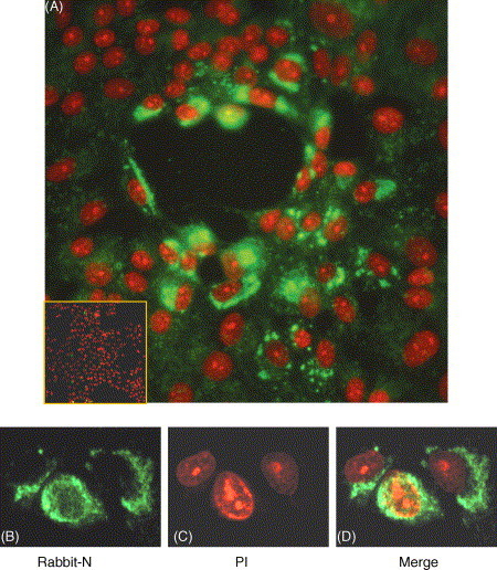
Detection of SARS–CoV N protein by indirect immunofluorescence assay in infected cells. Vero E6 cells were infected with SARS–CoV WHU strain. After 24 h cells were fixed and analyzed by indirect immunofluorescence using rabbit anti-N polyclonal antibody (green) ((A) and (B)) or pre-immune rabbit serum ((A) inset panel), followed by staining with PI (red) ((A) and (C)) to visualize the nuclear DNA. Differentially fluorescing images were gathered separately ((A) and (D)) using confocal microscope. Magnification 20 and 40× for (A) and (B)–(D), respectively.