
Fig. 3.
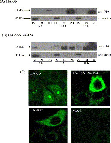
Cellular localization of HA-3b and HA-3bΔ124-154 proteins. Subcellular fractionation was performed to separate the cells into cytoplasmic (C), membrane (M) and nuclear (N) fractions. Western analyses were performed using with anti-HA (upper panel) and anti-actin (lower panel) monoclonal antibodies for each protein: (A) HA-3b; (B) HA-3bΔ124-154. The migration of protein molecular weight markers is indicated on the left. (C) The cellular localization of HA-3b, HA-3bΔ124-154, HA-Bax were analysed by indirect immunofluorescence at 18 h post-transfection. Mock transfected cells showed the specificity of the anti-HA monoclonal antibody.