
Fig. 5.
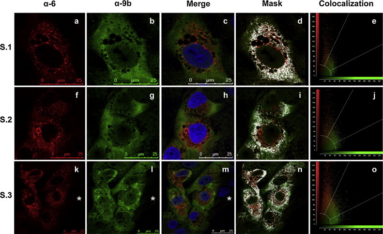
Proteins 6 and 9b partially co-localize in the cytoplasm of SARS-CoV infected cells. Vero E6 cell cultures were SARS-CoV infected and were fixed and analyzed by confocal immunofluorescence. Three representative examples are shown (S.1–S.2 and S.3). SARS-CoV protein 6 was detected with rat anti-protein 6 antibody and visualized with Tx-Red-conjugated anti-rat antibody (a, f, k). SARS-CoV protein 9b was detected with mouse anti-protein 9b antibody and visualized with FITC-conjugated anti-mouse antibody (b, g, i). Merge images show the localization of proteins 6, 9b, and DAPI stained nucleus in blue (c, h, m). The masks show the partial co-localization of proteins 6 and 9b in the cytoplasm of SARS-CoV infected cells (d, i, n). The graphics show the quantification of co-localization signal (e, j, o). The asterisk shows a non-infected cell, as a control. (For interpretation of the references to color in this figure legend, the reader is referred to the web version of the article.)