
Fig. 9.
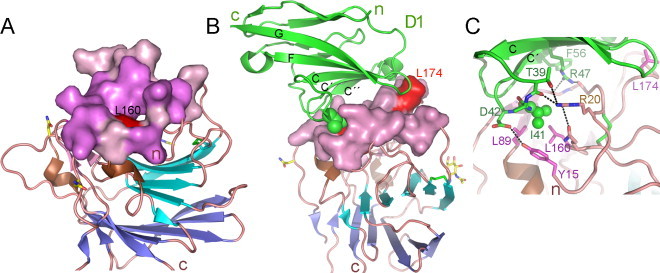
MHV recognition of its CEACAM1 receptor. (A) The MHV NTD structure with the CEACAM1-binding surface. The NTD ribbon diagram is shown as in Fig. 8B. The surface of the N-terminal MHV residues that form a socket is shown in violet and that of the other receptor-binding residues is pink. MHV Leu160 in the bottom of the socket is shown in red. (B) The MHV NTD in complex with the CEACAM1 receptor (PDB ID 3R4D) (Peng et al., 2011). The CEACAM1 N-terminal D1 is shown in green, with the β-strands in the receptor-binding CFG β-sheet labeled. The side chain of CEACAM1 Ile41 that penetrates the NTD socket is shown as spheres. The MHV Leu160 in the socket and Leu174 that contacts the top of D1 are in red. (C) Key virus–receptor binding motifs. Side chains of some receptor-binding MHV residues are shown, with carbons in pink; the hydrophobic residues in the bottom of the socket and Leu174 are in magenta; the CEACAM1 residues are in green. Ile41 in the CC′ loop, the most important virus-binding motif in CEACAM-1 (Peng et al., 2011), is shown as spheres.