
Fig. 1.
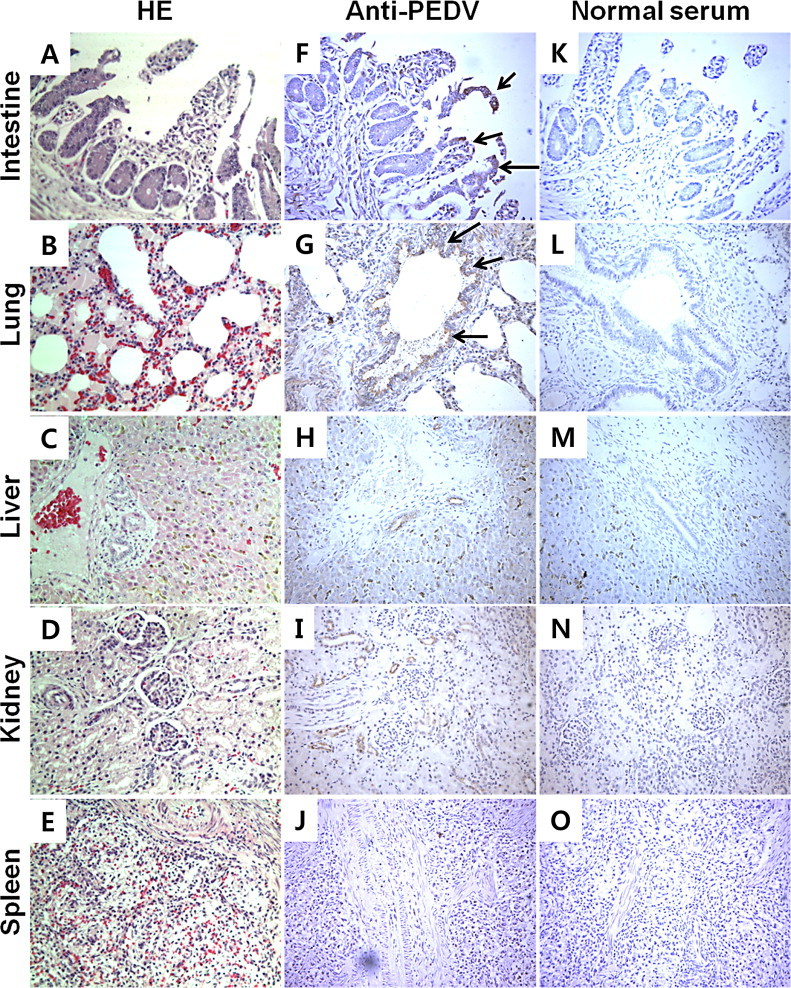
Pathological examination and IHC of samples from piglets naturally infected with PEDV. Samples from the small intestine, lung (bronchus and alveoli), liver, kidney and spleen stained with hematoxylin and eosin (HE) are shown in the left panel (A–E). Images from IHC with mouse polyclonal anti-PEDV antibody (F–J) or normal mouse serum (K–O) are shown in the middle and right panel, respectively. Arrows indicate PEDV-infected cells. Magnification: 100×.