
Fig. 4.
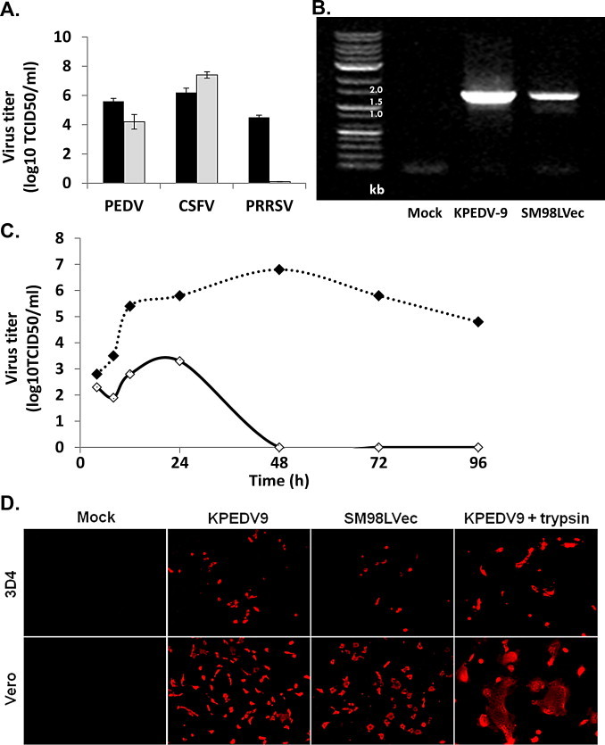
PEDV infection in porcine alveolar macrophages. (A) The susceptibility of 3D4 cells to PEDV. The 3D4 cells were infected with PEDV, CSFV (LOM strain), and PRRSV (VR-2332 strain), and progeny viruses were titrated in appropriate cell lines. Titers of 3 different swine viruses in the 3D4 cells (gray bar) were compared. For better understanding, viral titer was also compared with those in known susceptible cells (black bar); Vero cells for PEDV, PK15 cells for CSFV, and MARC145 cells for PRRSV, respectively. (B) The susceptibility of porcine primary alveolar macrophages to PEDV. Primary alveolar macrophages were collected and infected with Vero cell-adapted strains of PEDV and then virus infectivity was evaluated by RT-PCR. (C) Viral growth kinetics in alveolar macrophage cell line. Vero and 3D4 cells were inoculated with KPEDV9 and progeny viruses were titrated at the indicated time points. Vero (black circles) and 3D4 (white circles) cells were inoculated with Vero cell-adapted strains of PEDV and progeny viruses were titrated at the indicated time points. In Vero cells, the amount of KPEDV-9 increased continuously and gradually until reaching a titer of 107 TCID50 around 48 hpi, which slowly reduced afterwards. In 3D4 cells, the titer of KPEDV-9 increased slightly, reaching 103.4 TCID50 by 24 hpi. The titer was slowly decreased by 48 hpi, and we were unable to detect any infectious viruses after 48 hpi. (D) Vero and 3D4 cells were inoculated with Vero cell adapted PEDVs (KPEDV9 or SM98LVec) in supplemented with 10 μg/ml trypsin as indicated, and the cells were stained with mouse polyclonal anti-PEDV at 24 h after infection. Scale bar = 100 μm. A reduced infection in 3D4 cells was also supported by the results of an immunofluorescence assay. The percentage of PEDV-infected cells was lower in 3D4 cells than in Vero cells. We also compared CPE formation with trypsin condition.