Fig. 4.


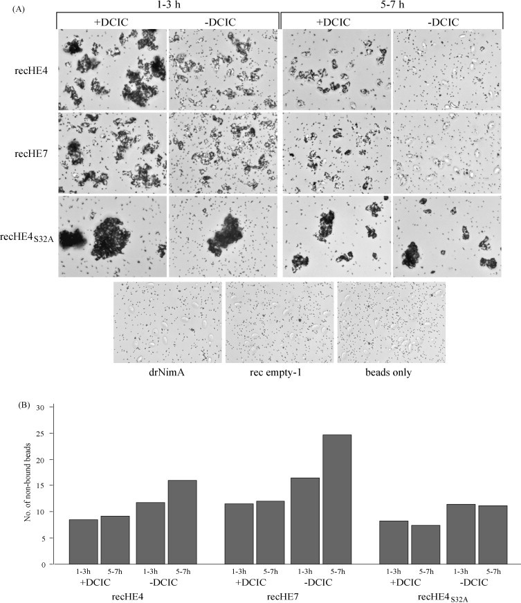
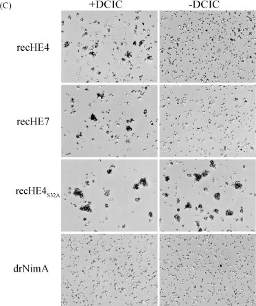
Agglutination of red blood cells (RBCs) with recHEs. RecHE4, recHE7 and recHE4S32A coupled to Dynabeads®TALON™ were pre-treated with DCIC in DMSO, or DMSO only, and mixed with RBCs. (A) The mixtures of Atlantic salmon RBCs (AsRBCs) and beads were observed under the microscope between 1–3 h and 5–7 h after adding AsRBCs. Negative controls included beads coupled with the His6-tagged protein drNimA, rec empty-1 and beads only. (B) Aliquots from the AsRBC-bead mixtures in (A) were taken out between 1–3 h and 5–7 h after addition of AsRBCs, and free beads (not associated with AsRBCs) were counted using a Bürker chamber. These experiments were repeated three times, and data from one representative experiment is presented. (C) The mixtures of rabbit RBCs (rRBCs) and beads were observed under the microscope 1 h after adding of rRBCs. Beads coupled with the His6-tagged protein drNimA served as a negative control. All photographs presented were taken at 40× magnification.
