
Fig. 5.
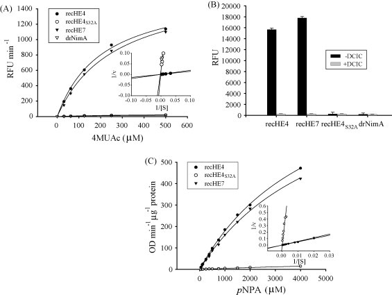
Acetylesterase activities of recHEs. (A) The acetylesterase activities of recHEs coupled to Dynabeads®TALON™ were determined using increasing concentrations of the substrate 4MUAc ([S]). The data (each data point is the average of at least three parallels) are presented by Michaelis–Menten and Lineweaver–Burk plots. The experiment was repeated twice, but only one is shown. Beads coupled to the His6-tagged protein drNimA served as negative control. RFU: relative fluorescence units; 1/v: reciprocal values of RFU min−1. (B) The influence of DCIC on acetylesterase activities of the recHEs coupled to Dynabeads®TALON™ using 4MUAc was studied. End-point data following 40 min incubation with substrate are shown. Beads coupled with the His6-tagged protein drNimA served as negative control. RFU: relative fluorescence units. (C) The acetylesterase activities of recHEs in solution were determined using increasing concentrations of the substrate pNPA ([S]). Rec empty-1 and drNimA served as negative controls (not shown). The data (each data point is the average of at least three parallels) are presented by Michaelis–Menten and Lineweaver–Burk plots. The experiment was repeated twice, but only one is shown. 1/v: reciprocal values of OD min−1 μg−1 protein.