
Fig. 6.
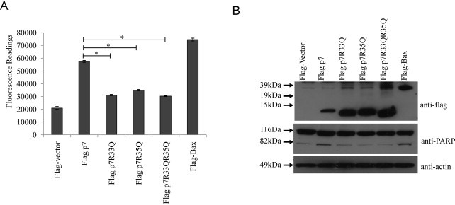
HCV p7 induced apoptosis is dependent on the cellular topology of p7 protein. (A) Apo-One fluorometric assay system from Promega Corporation was used to measure the activation of caspase-3/7 in Huh7.5 cells 24 h post-transfection with flag-vector, wild-type p7, substitution mutants (containing either R to A or changes) and the known apoptosis inducer, Bax. (B) Western blot analysis was performed to determine the expression levels of the different flag-tagged proteins (top) and the cleavage of endogenous PARP, which is a substrate of activated caspase-3, from 116 to 83 kDa (middle). Equal sample loading was verified by the detection of endogenous actin (bottom). p-Values indicated by asterisks are considered statistically significant, p < 0.01 (*).