
Fig. 8.
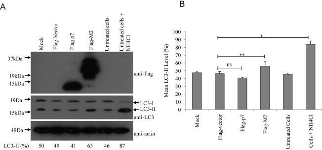
HCV p7 does not induce accumulation of autophagosomes. (A) Measurement of the conversion of LC3-I to LC3-II was used as an autophagic marker. 293FT cells, mock transfected, or transiently transfected with flag vector, flag p7, flag-M2 and untransfected cells untreated or treated with ammonium chloride were analyzed by Western blot analysis using anti-LC3 antibody (middle). Similarly, the expression levels of the different flag-tagged proteins were determined (top). Equal sample loading was verified by the detection of endogenous actin (bottom). The percentage conversion of LC3-I to LC3-II (i.e. LC3-II/LC3-I + LC3-II) is calculated after densitometry was used to quantify the intensities of LC3-I and II in the autoradiographs. Percentage LC3-II values are shown below the blots. (B) The mean LC3-II values quantified for four independent experiments were computed after densitometry and are plotted with standard deviations (error bars). p-Values indicated by asterisks are considered statistically significant, p < 0.05 (**), p < 0.01 (*) and no significant difference (ns).