
Fig. 9.
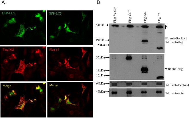
Interaction of M2 and p7 proteins with endogenous Beclin-1 and their effects on the accummulation of GFP-LC3 puncta. (A) Immunofluorescence analysis was performed on 293FT cells transfected with FLAG-tagged GFP-LC3 and expression plasmids encoding either FLAG-tagged M2 or p7. Twenty-four hours after transfection, cells expressing GFP-LC3 (green) and p7 or M2 (red) were analyzed by fluorescence microscopy. Merged images are shown below. (B) Co-immunoprecipitation of M2 and p7 proteins with endogenous Beclin-1. 293FT cells were transfected with flag-vector, flag-GST (negative control), flag-M2 and flag-p7. The cells were harvested at about 24 h post-transfection, lysed, and subjected to IP with anti-Beclin-1 antibody and protein A agarose beads. The amount of flag-tagged proteins that co-immunoprecipitated (IP) with Beclin-1 was determined by Western blot analysis (WB) with an anti-flag antibody (top). The amounts of flag-tagged proteins and endogenous Beclin-1 in the lysates before IP were determined by subjecting aliquots of the lysates to Western blot analysis (middle and bottom). The protein marked with Greek letter beta (β) represents the heavy chain of the antibody used for IP.