
Fig. 1.
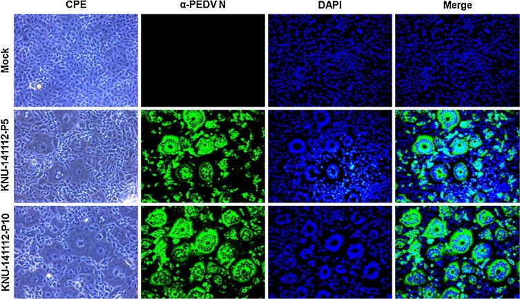
Cytopathology and IFA of a PEDV isolate KNU-141112 in infected Vero cells. Vero cells were mock infected or infected with PEDV KNU-141112-P5 and KNU-141112-P10 isolates. PEDV-specific CPEs were observed daily and were photographed at 24 hpi using an inverted microscope at a magnification of 200× (first panels). For immunostaining, infected cells were fixed at 24 hpi and incubated with MAb against the N protein, followed by Alexa green-conjugated goat anti-mouse secondary antibody (second panels). The cells were then counterstained with DAPI (third panels) and examined using a fluorescence microscope at 200× magnification. (For interpretation of the references to color in this figure legend, the reader is referred to the web version of this article.)