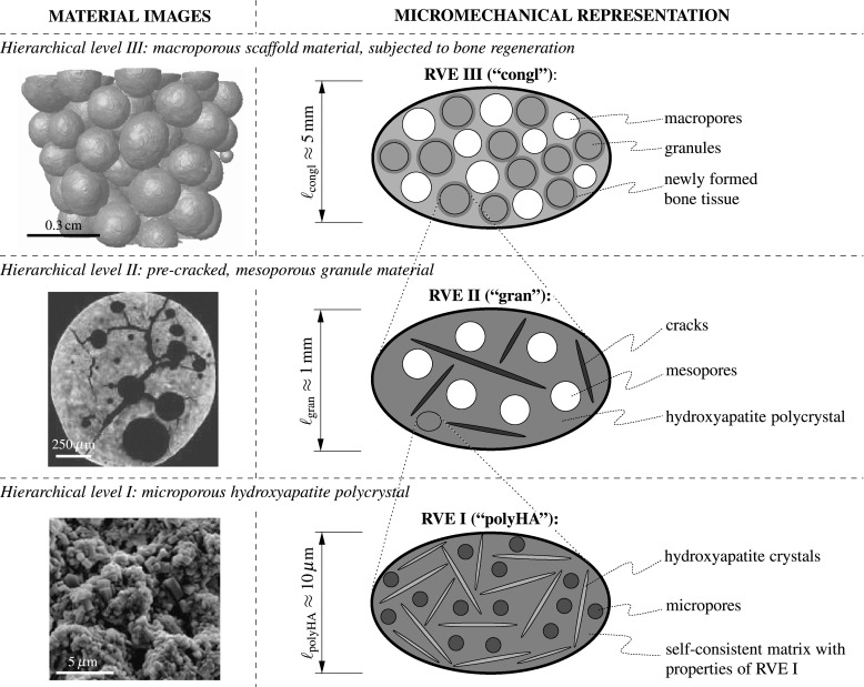
Fig. 1.

Three-level micromechanical representation of the hydroxyapatite-based granular biomaterial (column on the right-hand side), following the morphological features found in images on different observation scales (column on the left-hand side); the depicted images have been acquired by means of scanning electron microscopy (hierarchical level I) and CT imaging techniques (hierarchical levels II and III)
