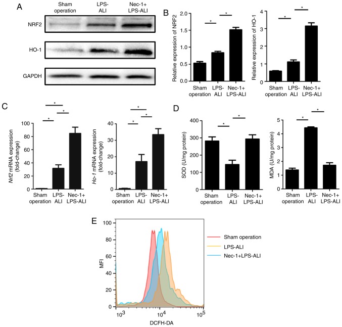
Figure 3.
Nec-1 attenuates the ROS response to LPS-induced ALI in vivo. (A) Western blotting of lysates in the indicated mouse lung tissues was used to estimate the expression levels of ROS-associated proteins (NRF2 and HO-1). (B) Semi-quantification of the protein expression levels of NRF2 and HO-1. Representative images of at least three independent experiments are shown. (C) mRNA expression levels of NRF2 and HO-1 in lung tissues from sham-operated, LPS-ALI and Nec-1 + LPS-ALI mice were detected by reverse transcription-quantitative PCR and were normalized to GAPDH. (D) Levels of SOD2 and MDA in the lung tissue homogenates were detected using commercial kits. (E) Intracellular ROS production of the indicated lung cells was analyzed by DCFH-DA staining and flow cytometry. Data are presented as the mean ± SEM from at least three independent experiments (n=5 mice/group). *P<0.05, as indicated. ALI, acute lung injury; HO-1, heme oxygenase-1; LPS, lipopolysaccharide; MDA, malondialdehyde; Nec-1, necrostatin-1; NRF2, nuclear factor erythroid 2-related factor 2; SOD2, superoxide dismutase.

