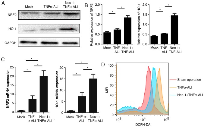
Figure 5.
Nec-1 regulates ROS in TNF-α-stimulated RLE-6TN cells. (A) Western blotting of lysates in the indicated alveolar epithelial RLE-6TN cells was used to detected the expression levels of ROS-associated proteins (NRF2 and HO-1). (B) Semi-quantification of the protein expression levels of NRF2 and HO-1. Representative images of at least three independent experiments are shown. (C) mRNA expression levels of NRF-2 and HO-1 in RLE-6TN cells from the mock, TNF-α-ALI and Nec-1 + TNF-α-ALI groups were detected by reverse transcription-quantitative PCR and were normalized to GAPDH. (D) Intracellular ROS generation was analyzed by DCFH-DA staining using flow cytometry. Data are presented as the mean ± SD from at least three independent experiments. *P<0.05, as indicated. ALI, acute lung injury; HO-1, heme oxygenase-1; Nec-1, necrostatin-1; NRF2, nuclear factor erythroid 2-related factor 2; ROS, reactive oxygen species; TNF-α, tumor necrosis factor-α.

