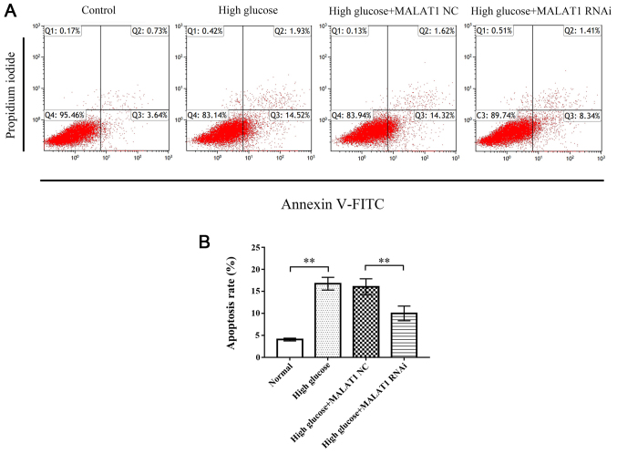
Figure 4.
Flow cytometry was used to detect CEP cell apoptosis. (A) Representative flow cytometric histograms. (B) The results revealed that the apoptosis rate of rat CEP cells treated with 25 mM glucose for 72 h was significantly increased. The apoptosis rate in the high glucose + MALAT RNAi group was significantly decreased compared with the high glucose and high glucose + MALAT1 NC groups. No change was observed between the high glucose and the high glucose + MALAT1 NC groups. **P<0.01 vs. the 5-mM glucose control group. CEP, cartilage endplate; MALAT1, metastasis associated lung adenocarcinoma transcript 1; NC, negative control.

