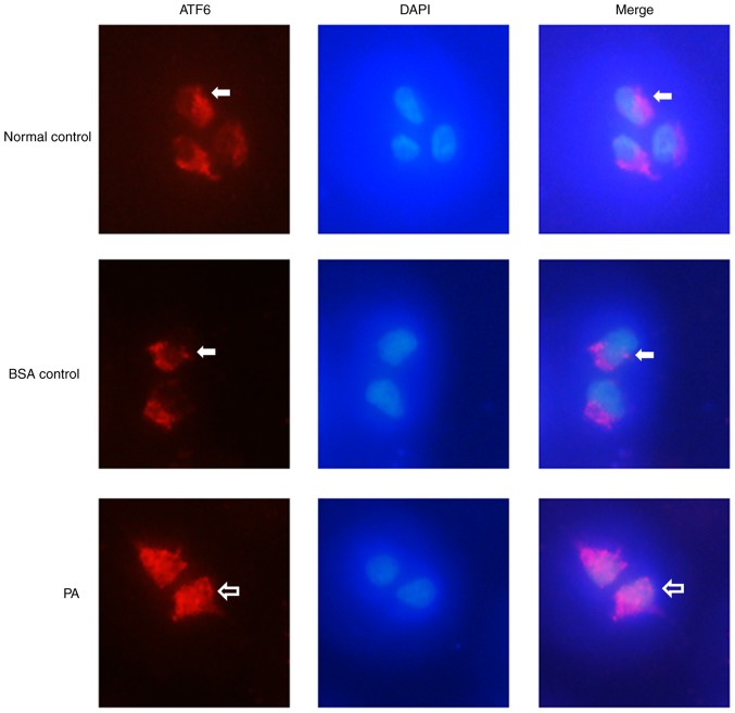
Figure 5.
Nuclear localization of ATF6 in INS-1 cells after PA treatment. ATF6 was visualized in red, DAPI nuclear staining in blue. ATF6 (white arrows) localized to the cytoplasm of INS-1 cells in the control group, with no ATF6 expression in the nucleus. Treatment with 0.5 mM PA induced nuclear localization of ATF6 in INS-1 cells; ATF6 was expressed in both the cytoplasm and the nucleus (blank arrows). Magnification, ×40. ATF6, activating transcription factor 6; PA, palmitate.

