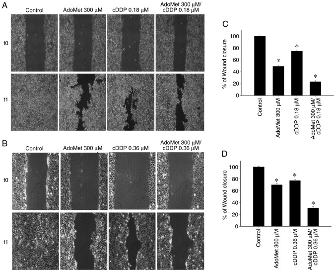
Figure 6.
Effect of AdoMet/cDDP combination on cellular migration of HNSCC. The migratory ability of (A) Cal-33 and (B) JHU-SCC-011 cells was measured by wound healing assay. Confluent monolayers of Cal-33 and JHU-SCC-011 cells treated or not (control), with 300 µM AdoMet with/without 0.18 µM cDDP for 24 h or 0.36 µM cDDP for 48 h, respectively, in comparison to treatment with cDDP alone were scratched with a micropipette tip and snapshot images were captured using microscope to examine for wound closure. Images of the wounds corresponding to time zero (T0) and after 24 h (T1) of scraping in both cell lines are presented. (C and D) Histograms reporting the quantification of the wound area calculated as a percentage of the control using ImageJ software are depicted. Data represent the average of 3 independent experiments. The means ± SD are shown. *P<0.05 vs. control. HNSCC, head and neck squamous cell carcinoma; AdoMet, S-adenosyl-L-methionine; cDDP, cisplatin.

