
Fig. 1.
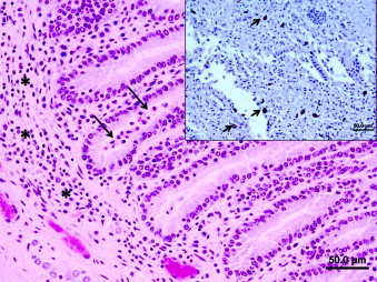
Small intestine (jejunum); scouring calf: there are low to moderate numbers of lymphoplasmacytic cells along with rare eosinophils that infiltrate the submucosa and lamina propria (black asterisks). Occasional crypts are distended by low numbers of cellular debris (long black arrows). H&E, bar = 100 μm. Inset: many intact Giardia trophozoites (short black arrows) are present within the sloughed intraluminal cellular debris. IHC, bar = 50 μm.