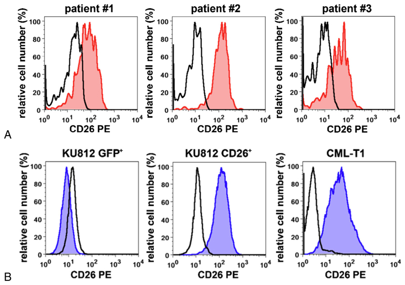
Figure 1. Expression of DPPIV (CD26) on the surface of CML cells.
(A) CD34+/CD38− LSCs from three patients with CML (patients #1, #2, and #3 in Supplementary Table E2, online only, available at www.exphem.org) were examined for expression of CD26 (red histograms) by multicolor flow cytometry. The staining reaction obtained with an isotype-matched control antibody is also shown (open black histograms). (B) Expression of CD26 on KU812 cells transduced with a control construct (KU812 GFP+) or human CD26 (KU812 CD26+) and on the CD26+ CML-T1 cell line. Expression of CD26 was assessed by flow cytometry, as described in the text. Blue histograms represent expression of CD26 and the black open histograms indicate staining reactions obtained with an isotype-matched control antibody.

