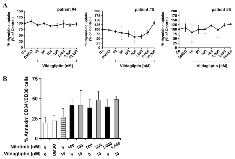
Figure 3. In vitro effects of vildagliptin alone or in combination with imatinib or nilotinib on growth and survival of primary CML SCs (LSCs).
(A) Primary CML MNCs were incubated in control medium (Co) or in various concentrations of vildagliptin (10–10,000 nmol/L) at 37°C for 48 hours and then 3H-thymidine uptake was measured. Results are expressed as a percentage of control and represent the mean ± SD from triplicates. The dimethyl sulfoxide (DMSO) control is also shown. Patient numbers (#) refer to Supplementary Table E2 (online only, available at www.exphem.org). (B) CML MNCs were incubated in control medium (0) or in nilotinib (100–1,000 nmol/L) or vildagliptin (10 μmol/L) alone or in a combination at 37°C for 48 hours. After incubation, cells were stained with antibodies against CD34, CD45, CD38, and Annexin V to determine apoptosis in CML LSCs. DAPI was used as a viability marker to exclude nonviable cells. White bars represent medium control (0) or dimethyl sulfoxide (DMSO) control; the lined bar in grey represents cells incubated with vildagliptin alone; black bars represent cells incubated with nilotinib alone; and grey bars represent apoptosis induction after incubation with both drugs. Results are expressed as a percentage of Annexin-positive CD34+/CD38− cells and represent the mean ± SD from three independent experiments (patients #6, #7, and #8).

