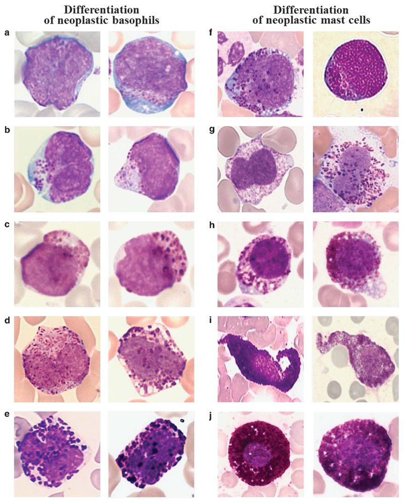
Figure 1. Morphology of basophils and mast cells and their precursor cells.
Morphological changes typically occurring during the differentiation of neoplastic basophils (left panels) and mast cells (right panels). (a–e) Basophil-lineage cells were examined on Wright–Giemsa-stained PB smears in patients with CML at the time of acceleration and basophil expansion (a–c) or chronic phase CML with moderate basophilia (d, e). (a) Two metachromatically granulated blast cells. (b) Two basophilic promyelocytes. (c) Two basophilic myelocytes. (d) Two immature basophils with bi-lobed nuclei, and (e) two fully mature basophils with segmented nuclei. (f–j) Mast cell lineage cells on Wright–Giemsa-stained BM smears of a patient with mast cell leukemia (f–h) and a patient with indolent systemic mastocytosis (i, j) are shown. (f) Metachromatic blast cells. (g) Atypical mast cells type II (promastocytes) with bi- or poly-lobed nuclei. (h, i) Immature and more mature atypical mast cells type I with cytoplasmic extensions and hypogranulated cytoplasm, and (j) mature typical round mast cells with a round central nucleus. Note that it is impossible to distinguish between mast cells and basophils by morphology at the stage of metachromatic blasts. Images were prepared using a Basler Vision Technology A1021C camera (Ahrensburg, Germany) connected to an Olympus BX51 microscope equipped with Olympus UIS 2 U Plan FL N × 100/1.30 objective lens (Olympus, Hamburg, Germany). Images were prepared using TRIBVN ICS capture 1.6 and processed with PowerPoint software (Microsoft, Redmond, WA, USA). Original magnification: × 100.

