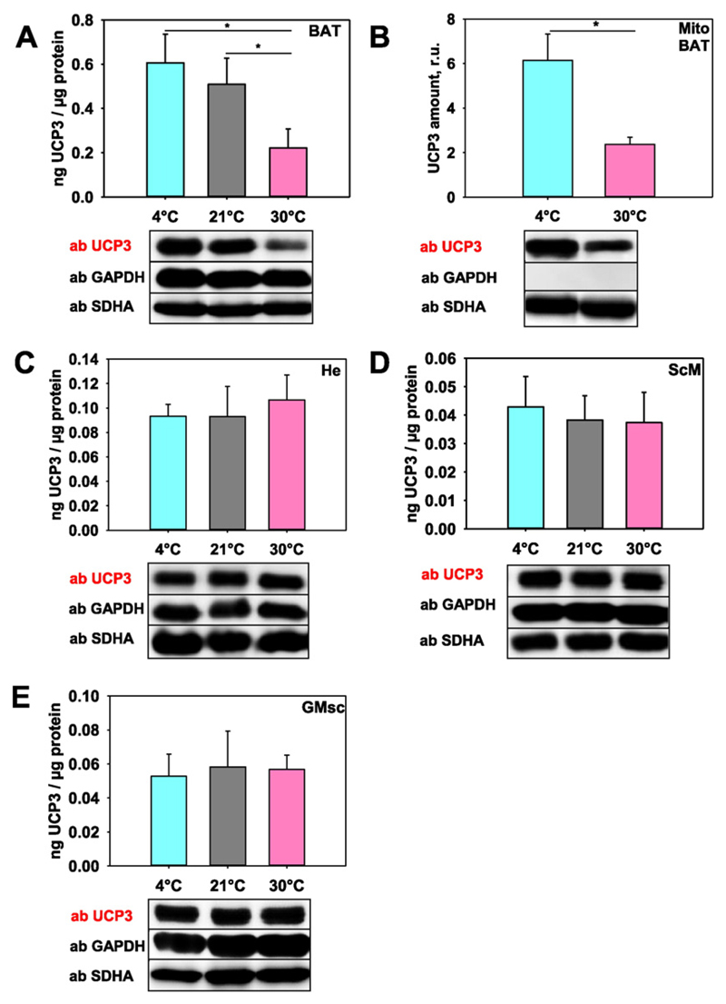
Fig. 3. Quantification of UCP3 expression at different temperatures.
(A) UCP3 amount in tissue lysates from brown adipose tissue, (C) heart, (D–E) skeletal muscles (ScM, D; GMsc, E) and (B) mitochondria isolated from BAT. Wild type mice were housed at 30 °C, 21 °C and 4 °C. 20 μg of total protein from tissue or 10 μg isolated mitochondria were loaded per lane. The relative UCP3 amount (r.u.) is a ratio between the band intensity of standard (mUCP3, 5 ng) and sample. Five mice were tested at 30 °C and 4 °C and four mice at 21 °C.

