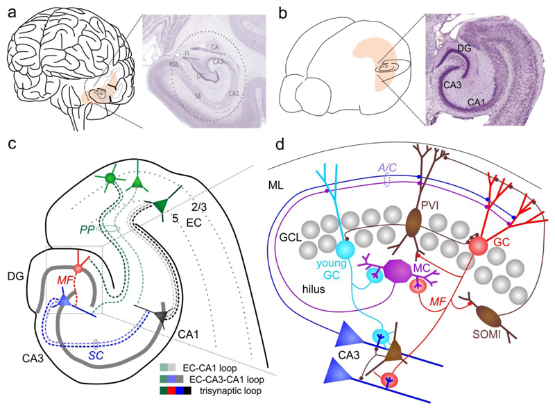
Fig. 1. Anatomical organization of the hippocampus.
a | Human brain and Nissl-stained section through the hippocampus. b | Same as in a for a mouse brain. c | Schematic of the mouse hippocampal formation and its main synaptic connections. d| Illustration of the DG microcircuitry, its intrinsic connections and outputs to CA3. A/C, associational-/commissural pathway; DG, dentate gyrus; EC, entorhinal cortex; GC, granule cell; GCL, granule cell layer; MC, mossy cell; MF, mossy fiber; ML, molecular layer; PP, perforant-path; PVI, parvalbumin-expressing interneuron; SC, schaffer-collateral; SOMI, somatostatin-positive interneuron. Photographs in a and b are derived from www.brainmaps.org and Franklin, K. B. J. & Paxinos, G., ‘The mouse brain in stereotaxic coordinates’, 3rd edition (Elsevier, New York, 2007), respectively.

