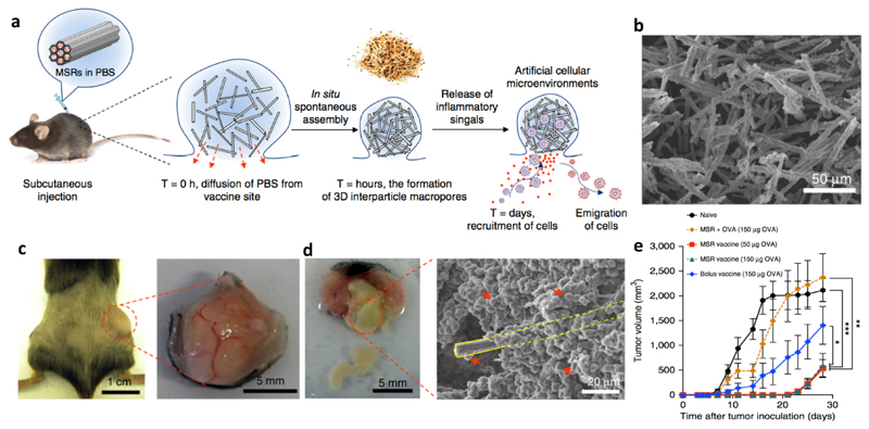
Figure 4. Injectable MSRs recruiting antigen-presenting cells.
a: Schematic representation of the working mechanism of the vaccine. MSRs formed a local nodule after subcutaneous injection to recruit APCs which were matured, loaded with antigens, and emigrated to dLN to generate effector T cells. b: Scanning electron microscope image of the MSRs, which formed a nodule after subcutaneous injection in mice (c) and elicited cell infiltration in the nodule (d). The yellow rectangle in panel d marks one MSR, which is surrounded by cells as indicated by the red arrows. This vaccine efficiently inhibited the growth of EG.7-OVA tumors in mice (e). Adapted from ref. 70, with permission from Springer Nature, copyright 2015.

