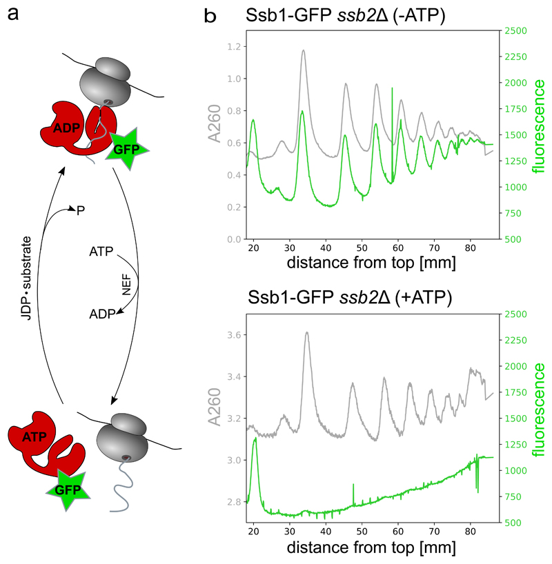
Figure 3. Detected Ssb-GFP in the sucrose gradient reflects the association of the Hsp70 to ribosomes.
(a) Hsp70 cycle for the example of Ssb-GFP binding to ribosomes. (JDP = J-domain protein, NEF = nucleotide exchange factor)
(b) Polysome profiles recording the co-migration of Ssb-GFP with ribosomes. Ribosome and Ssb-GFP co-migration is analyzed by simultaneous detection of A260 (grey) and GFP fluorescence (green) using the TRIAX™ flow cell (BIOCOMP instruments). Ssb-GFP cell lysate was either thawed in the presence of hexokinase and 0.2% glucose for rapid ATP depletion (-ATP, upper panel) or in the presence of 1 mM ATP (+ATP, lower panel).

