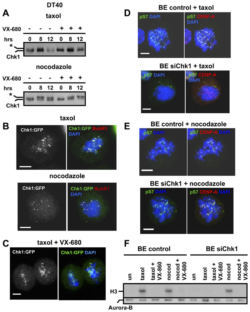
Figure 5. Chk1-Deficient Cells Exhibit Decreased Aurora Activity during Treatment with Taxol.
(A) Mitotic phosphorylation of Chk1 does not require Aurora activity. Western blot analysis of total Chk1 during treatment of G2/M-elutriated DT40 cells with taxol or nocodazole, in the absence or presence of VX-680. The asterisk marks phosphorylated Chk1.
(B and C) Localization of Chk1 to kinetochores is not dependent on Aurora activity. Chk1−/− cells expressing Chk1:GFP were treated with taxol or nocodazole for 2 hr in the (B) absence or (C) presence of VX-680 and were analyzed by confocal microscopy. Red: BubR1; green, Chk1:GFP; blue, DNA. A single image plane is shown. The scale bar is 5μm
(D and E) Projected deconvolved image stacks of BE cells transfected with negative siRNA (control) or with Chk1 siRNA(siChk1) and treated with (D) taxol or (E) nocodazole for 2 hr. Red, CENP-A; green, phosphorylated Serine 7 of CENP-A (pS7); blue, DNA. The scale bar is 5 mm.
(F) BE cells transfected as in (D) were treated with taxol, nocodazole (nocod), or VX-680 for 6 hr. Upper panel: Aurora-B-associated histone H3 kinase activity. Lower panel: western blot analysis of immunoprecipitated Aurora-B.

