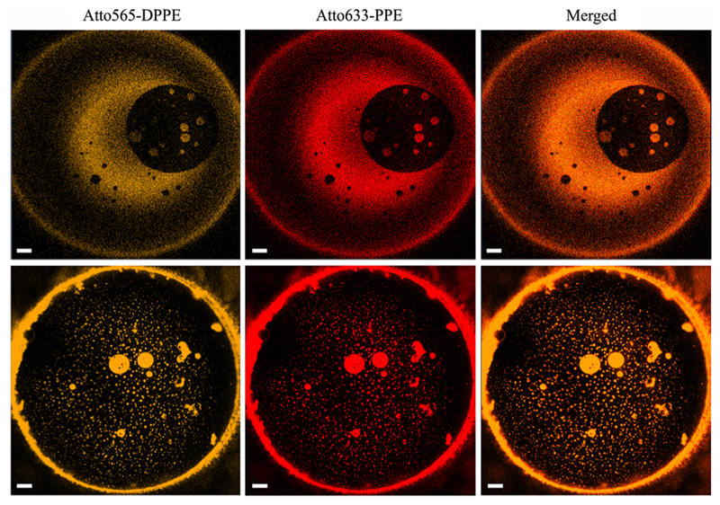
Fig. 1.
Domains of all sizes from the two membrane leafs are in register. The bright spots from the two monolayers always coincide: the left column displays micrographs that were obtained by exciting Atto565–DPPE in the cis monolayer; the middle column shows the fluorescence of Atto633–PPE in the trans monolayer of the same membrane at the same time; the right column shows perfect overlap of both channels. The upper and lower rows were obtained in two subsequent experiments at room temperature (T = 295 K). The lipid composition was diphytanoyl phosphatidylcholine (DPhPC): dipalmitoyl phosphatidyl choline (DPPC): photoswitchable diacylglycerol (PhoDAG–1): cholesterol 2:1:1:2 plus 0.004 mol% Atto565–DPPE in the cis monolayer and 0.004 mol% of Atto633-PPE in the trans monolayer. The buffer contained 20 mM HEPES and 20 mM KCl (pH = 7.0). The scale bar has a length of 20 μm.

